[tab_nav type=”two-up”][tab_nav_item title=”Clinical Case” active=”true”][tab_nav_item title=”Answer” active=””][/tab_nav][tabs][tab active=”true”]
As you sit in the ICU watching the latest lecture on MarylandCCProject you get called to assess your newest transfer from the floor. When you arrive to bed 7 you see a 57 y/o Hispanic woman s/p orthotopic liver transplant (a byproduct of long term Hep C cirrhosis) was transferred to the ICU from the transplant medicine floor for encephalopathy of unclear etiology.
The patient had experienced several months of subacute decline in mental status and on admission was found to have persistant coagulase negative staph bacteremia concerning for infectious endocarditis. Her ICU course was prolonged and complicated by respiratory failure requiring mechanical ventilation. Her husband eventually decided not to escalate care and the patient was placed on comfort measures, but was not extubated.
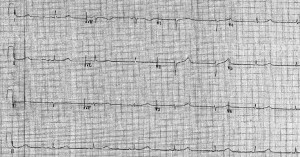
During the final week of her life, her temperature, heart rate and blood pressure slowly decreased. A very eager intern performed an ECG when her temperature was 31.7C.
What do you notice?

Hint: Focus on the lead II/rhythm strip…….
What do you see? What would be your next step?
[/tab][tab]
J waves or “Osborn” waves; A normal finding in hypothermia, no additional treatment needed!
J waves (also known as “Osborn” waves) were first described on the ECG in 1938 by Tomaszewski and, subsequently, by several other researchers, most famously by J.J. Osborn in his now classic 1953 paper in the American Journal of Physiology. They are so-named because they occur at the J point that connects the S wave to the S-T segment.
[Little known is that the nomenclature “J wave” was accepted only after multiple other suggestions, including “Jay-Z,” “J. Crew,” “alt-J” and “Jumpin’ Jehosawave” all did not stick—NB: I most likely fabricated this information].
Classically, J waves are associated with:
- extreme hypothermia
- hypercalcemia
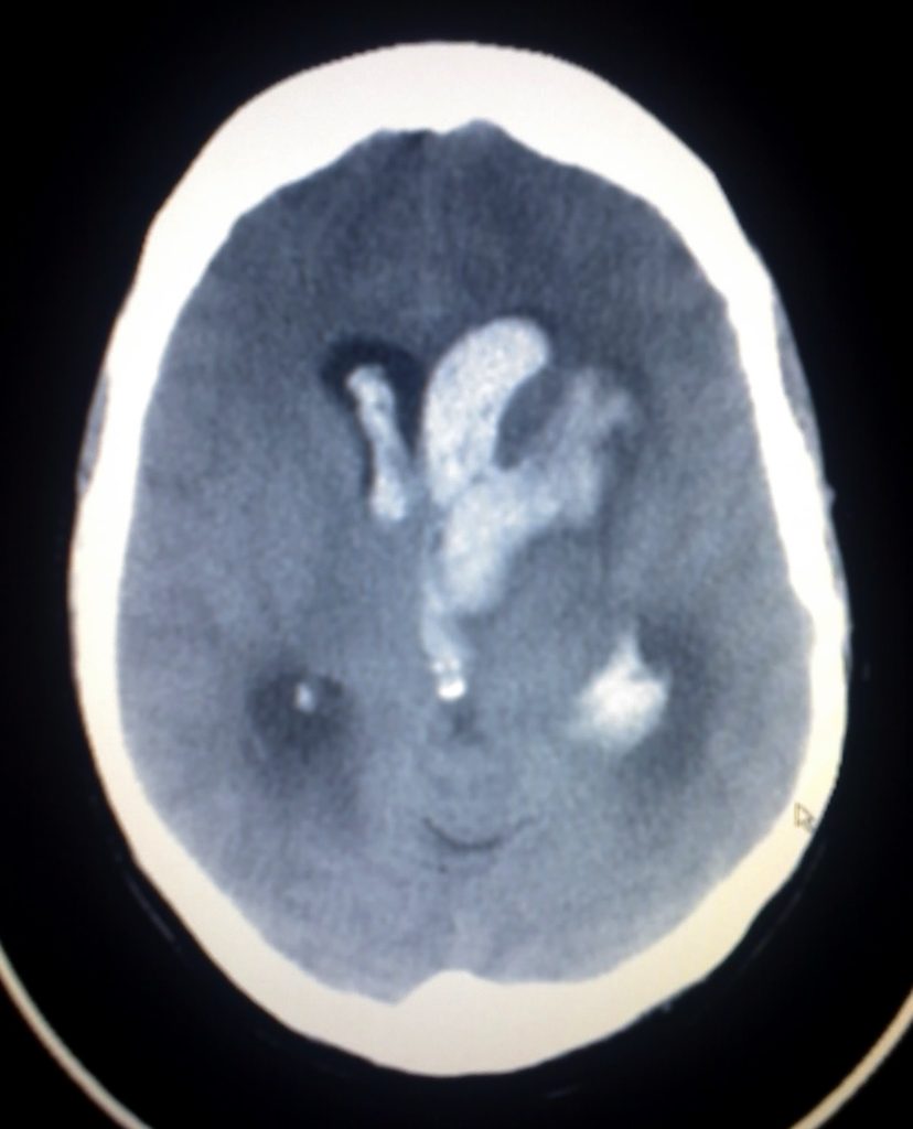
- traumatic or hemorrhagic brain injury.
They are thought to develop from a temperature-modulated increase in epicardial transient outward currents that lead to altered action potentials in the epicardium and manifest on the surface ECG as J waves.
Other common hypothermia-related ECG changes include QTc prolongation and bradycardia, both of which are seen here (QT/QTc = 624/527ms; HR = 43 bpm).
Treatment: Therapy is aimed at correction of the cause of the J wave.When related to hypothermia its best to rewarm the patient. The J wave won’t kill the patient, but the inevitable Ventricular arrhythmias associated with hypothermia will…………….
Reference
- Osborn, J.J. “Experimental hypothermia; respiratory and blood pH changes in relation to cardiac function.” Am J Physiol (1953). Dec;175(3):389-98.
- Gussak I et al. “ECG phenomenon called the J wave. History, pathophysiology, and clinical significance.” J Electrocardiol (1995). Jan;28(1):49-58.
- Hurst JW. “Naming of the waves in the ECG, with a brief account of their genesis.” Circulation (1998). Nov 3;98(18):1937-42
[/tab][/tabs]


