Thanks to Drs. Katelyn Donohue and Christina Tupe for this fantastic Case Report!
After a long night of all-out chaos you hear of a transfer accepted by the previous shift and fast approaching. You grab a cup of coffee as even the seasoned charge nurse seems concerned over this one:
“A 45 yo AAF with a PMH of lupus nephritis and a previous episode of myocarditis is transferring to you ASAP for eval of ECMO/VAD to treat severe cardiogenic shock. Two days ago she developed acute shortness of breath at home and on arrival the OSH ER she was found to be severely hypoxic and required immediate intubation. During induction she PEA arrested for 90 seconds but achieved ROSC. Her oxygenation requirements increased to 100% FIO2 of and she required proning. Additionally, she has been persistently hypotensive requiring high doses of levophed, vasopressin, and epinephrine. A bedside stat echo showed an severely depressed EF (<15%).”
Thankfully the rest of the team will not arrive for another 3 hours, so this fun is coming just for you!
On arrival her respiratory and hemodynamic status have improved, but as expected she is in severe multi-organ failure. She has a profound metabolic acidosis, acute renal failure, transamonitis, hemolytic anemia and thrombocytopenia. Her fibrinogen is >600 and Ferritin is >5x upper limit of normal.You have this diagnosis: Sepsis, sepsis, and sepsis…. then notice her right arm!
The nurse takes off the tape, thinking it may be an allergic reaction and finds:
 You run over to her right side and determined that you cannot obtain a pulse in the right wrist at any point below the antecubital fossa. STAT duplex imaging shows clots in the following vessels: Right common femoral vein, right basilic and cephalic veins, left cephalic vein, right brachial right axillary vein, right radial and ulnar arteries.
Additionally, the nurse states that the patient is having bloody secretions from her ET tube and a quick bedside bronchoscopy shows signs of diffuse alveolar hemorrhage.
You grab the US, repeat an echocardiogram, and find an EF of 25% with severe hypokinesis, tricuspid regurgitation, and pulmonary hypertension with a moderately dilated inferior vena cava (but we are still on high dose Epinephrine, so what did you expect!?)
As you walk back to the office you scan the back pages of the transfer document and notice that oddly the following labs were sent and were all found to be negative: DS DNA, Cardiolipin, Beta 2 glycoprotein, Phosphatidylserine, Smith AB, Myeloperoxidase AB, SS-A, SS-B, Atypical pANCA, Neutrophil Cytoplasmic Antibody, HIT Ab, Serotonin release assay, Viral Panel, Blood cultures.
What is the diagnosis? What treatment measures should be started immediately to stabilize this patient?….. [expand title=” Think it over and then click here…” swaptitle=” “] Answer:
Antiphospholipid Syndrome (APS): autoimmune condition with vascular thrombosis and/or pregnancy morbidity with antiphospholipid antibodies (aPL).
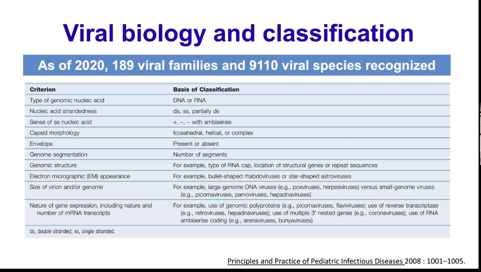
Catastrophic Antiphospholipid Syndrome (CAPS) a potentially life-threatening variant with small and large vessel thrombosis causing multiorgan failure (aka: Asherson’s syndrome)Diagnostic criteria for CAPS
Pathogenesis
- APS has a multifactorial pathogenesis involving both innate and adaptive immunity
- Theoretical genetic predisposition – thrombosis and antiphospholipid (antiPL) antibody production
- Transition to CAPS requires a “second hit” (often infectious) to incite the inflammatory reaction.
- “Cytokine storm” causes the acute thrombosis via a procoagulant effect
- New research is suggesting that antiPL antibodies cause endothelial damage via toll-like receptors and induce vasculopathy.
- Triggering factors:
- Infections (46%)
- Neoplasms (17%)
- Trauma/Surgery (16%)
- Anticoagulation problems (low INR or anticoagulant drug withdrawal) (10%)
- Medications (7%)
Demographics
- 72% women
- Mean age: 37yo
- 58% have primary APS, 27% have SLE, 5% have a lupus-like disease, 9% have another autoimmune disease
Clinical Features
- Symptoms depend on the extent of organ thrombosis and and manifestations of the SIRS reaction:
Diagnosis
- Lupus Anticoagulant: present in 70-80% of cases; can cause both false neg and false positive results
- IgG anticardiolipin (aCL): present in 80% of cases.
- IgM aCL: present in 48% of cases.
- B-2GPI IgG and IgM: 11.1% and 3.2% of cases, respectively
- “Seronegative CAPS” may be a result of consumption of antibodies during the acute phase of disease
- Ferritin: massively elevation; differential diagnosis: sepsis, CAPS, Stills disease and Macrophage Activation Syndrome.
- Acts as an pro-inflammatory signaling molecule beyond being an acute phase reactant.
- The precise role of Ferritin elevation in CAPS is not known, the presence of hyperferritinemia (present in 70% of CAPS) is a useful in aiding diagnosis
- Thrombocytopenia: present in about 60% of cases and usually associated with microangipathic hemolytic anemia
Differential Diagnosis
Treatment
- Early recognition and treatment is imperative to preventing mortality
- #1 goal: Control the inflammatory response and manage the thrombotic complications.
- Patients given combined therapy of AC, steroids, plasma exchange and IVIG have improved outcomes compared to patients who were not given plasma exchange or IVIG
- First-line therapy:
- Anticoagulation: heparin is mainstay of treatment by inhibiting clot generation, promoting fibrinolysis and possibly preventing aPL antibodies from binding to targets.
- Corticosteroids: minimum 3 days (1000mg of methylprednisone for 3-5 days)
- Be aware that steroids alone have not been shown to improve outcomes.
- Second-line therapy:
- Plasma exchange: improves patient survival in a registry of patients with CAPs
- Emphasized importance in patient with microangiopathic hemolytic anemia
- Remove 2-3 L or plasma for minimum 3-5 daysIVIg 0.4 g/d/kg for 4-5 days; may be helpful in patients with severe thrombocytopenia
- Plasma exchange: improves patient survival in a registry of patients with CAPs
- Third line therapies:
- Cyclophosphamide: may be beneficial in patients with SLE for immunosuppression
- Rituximab: This is the most studied biologic used in treatment of CAPS and has only been reported as being administered in about 20 patients with inconclusive results.
Prognosis
- 30% mortality
- Factors associated with poor outcomes: >36yo, pulmonary and renal involvement, active SLE, positive ANA
Back to your lady:
She was anticoagulated with Argatroban until HIT was effectively ruled out. She improved significantly with anticoagulation, high dose steroids and dialysis. She was extubated without event and her EF improved to 35% in 5 days. She continued to have vascular thrombosis events, and will need anticoagulation for life. Debridement was needed of the UE, but no amputation was required due to early diagnosis and therapy.
References
- Aguiar, C and Erkan, D. Catastrophic antiphospholipid syndrome: how to diagnose a rare but highly fatal disease. Ther Adv Musculoskelet Dis. Dec 2013; 5(6): 305–314.
- Cervera R, et al. 14th International Congress on Antiphospholipid Antibodies Task Force Report on Catastrophic Antiphospholipid Syndrome. Autoimmun Rev. 2014 Jul;13(7):699-707.
- Cervera R. Update on the diagnosis, treatment, and prognosis of the catastrophic antiphospholipid syndrome. Curr Rheumatol Rep. Jan 2010; 12: 70-76.
- Espinosa G, Cervera R, Asherson RA. Catastrophic antiphospholipid syndrome and sepsis. A common link? Jour of Rheum. 2007; 34(5): 923-926.
- Rosário C, et al. The hyperferritinemic syndrome: macrophage activation syndrome, Still’s disease, septic shock and catastrophic antiphospholipid syndrome. BMC Med. 2013 Aug 22;11:185.
[/expand]


