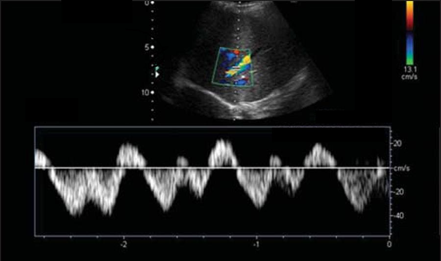
You are sitting in the call room, counting down the minutes until you are finished covering the critical care floor when you get a phone call from one of the floor surgical teams: “We are super busy and we have a paracentesis that needs to be done…. do you have any time to come help us out?..”
You check the INR and platelets and can’t find a logical reason to refuse. You finally ask for a background story:
“The pt is a 52 yo F with chronic liver failure due to alcoholism. Pt has been on the surgical service for the last week because of a recurrent pancreatic pseudocyst that required surgical management (POD #4). Pt has been doing well but her continued abdominal distension is starting to cause diffuse, mild abdominal pain and a slight rise in her creatinine. The pt had been on a fairly consistent paracentesis schedule, requiring a tap every 10 days for relief of pressure. The pt is currently awaiting a liver/pancreas transplant. Pt is otherwise in fine condition and may even be discharged home in the next 48 hours.”
You grab supplies, including a few vacuum bottles, and head off to the floor. You go through the motions of using the US to find a pocket, clean the skin, and pass an 18G angiocath into the peritoneal space without incident. You are quite surprised when this is what come out:
You note that her abdomen isn’t very tender, she has no leukocytosis or fever, and she otherwise looks and feels amazing. You send off labs showing: cell count 1670 (85% lymphocytes), total protein: 3.7 g/dl, glucose: 45 mg/dL….. The Gram stain and culture is negative so far (<24 hours). You are worried about an infection and recommend prophylactic SBP antibiotics, but you think you might be missing a more important diagnosis….
What diagnosis are you worried about? What labs could you send to assist in obtaining this diagnosis? What process could be causing this cloudy fluid collection? What treatment measures should be started immediately to stabilize this patient?….. [expand title=” Think it over and then click here…” swaptitle=” “]
Answer: Chylous Ascites (CA)
- An uncommon form of ascites (incidence: 1 in 11,589) caused by damage or obstruction to the lymphatic system → triglyceride rich ascitic fluid
- Higher risk with pt’s possessing limited reserve of anastomotic channels are at greater risk
- Present in ~1% of all cirrhotic patients
- Mortality: overall 71% (90% when related to cancer, 43% when related to trauma)
- Causes:
- Abdominal malignancies (25%, with 1/3 due to lymphoma) and cirrhosis (16%)– most common in developed countries (responsible for 2/3 of all cases)
- Chronic infections are most common in developing countries (Tb, filariasis)
- Pancreatitis is becoming a common cause of CA due to both compression of lymphatic channels and direct damage by pancreatic enzymes
- Abdominal malignancies (25%, with 1/3 due to lymphoma) and cirrhosis (16%)– most common in developed countries (responsible for 2/3 of all cases)
Said A. Chylous Ascites: Evaluation and Management
- Pathophysiology based on 3 basic mechanisms:
- Lymph exudation through walls of retroperitoneal megalymphatics (w/ or w/o a fistula)
- Leakage of lymph from the dilated subserosal lymphatics on the bowel wall (malignant infiltration of lymph nodes leading to blockade)
- Direct leakage of lymph through a lymphoperitoneal fistula associated with retroperitoneal megalympatics due to disruption associated with trauma or surgery
- Additionally worsened by:
- Increased caval and hepatic venous pressures assoc with constrictive pericarditis, right sided heart failure, and dilated cardiomyopathy → hepatic congestion → large increases in lymph production
- Cirrhosis → increased formation of hepatic lymph (treated with portal venous decompression)
- Complications
- Loss of essential proteins, lipids, immunoglobulins, vitamins, electrolytes, and water → severe immunopuppression and malnutrition
- Loss of drug bioavailability (dig, amio, cyclosporin)
- Evaluation/Diagnosis
- Clinical– painless abdominal distension (81%) or non-specific abdominal pain (14%), can be more acute s/p surgery, can also have wt loss, anorexia, malaise, steatorrhea, malnutrition, fevers, night sweats, or LAN
- Laboratory– by paracentesis:
- Must distinguish from psuedochylous ascites: has a similar appearance (due to cellular degeneration from infection or malignancy) without high TG level
 A. Cárdenas and S. Chopra- Chylous ascites
- Imaging:
- CT: eval for fluid (easily distinguished from blood), a fluid-fluid level, LAN, or masses
- Lymphoscintigraphy: follow lymph drainage, benign test but technically difficult
- Lymphangiography: GOLD standard to eval for blockage, can also treat resistant leaks, numerous complications (nephropathy, fat embolism, necrosis) make it unpopular
- Surgery (laparoscopy or laparotomy): to both diagnose and treat

- Treatment
- #1: treatment of the underlying cause!
- Diet: high-protein, low-fat diet with medium chain TGs (will be directly absorbed into intestinal cells) with long chain TG restriction = less chyle production; TPN can also help as it bypasses the gut TG absorption
- Pharm:
- Somatostatin/Octreotide + TPN can lead to near complete resolution (inhibits lymph excretion by acting at intestinal wall receptors) and can close fistulas
- Orlistat (a reversible inhibitor of gastric and pancreatic lipase) will minimize TG levels in ascites
- Etilefrine (a sympathomimetic) can resolve post-operative chylous effusions
- Therapeutic paracentesis without albumin replacement (unless cirrhosis is present)
- TIPS for cirrhosis, angiography with embolization for post-op CA
- Surgery:
- Important for post-op, neoplastic, or congenital cases
- Options include fistula closure, bowel resection, or insertion of a peritovenous shunt
Case resolution: You send off additional labs on the cloudy ascites and find that the TG level is 472, the amylase is <30, and there was no sign of Tb or other infectious causes. Due to the continued chylous collection the surgical team took the pt back to the OR. On a re-exploration of the abdominal cavity by laparotomy a lymphoperitoneal fistula was found and repaired. It was a worrisome complication of her pseudocyst resection and something that was also a known complication of her pancreatitis (though not likely the primary cause for this patient as the amylase was very low). The pt also underwent a detailed evaluation for a malignant source for the CA, which was entirely negative. The pt was sent home and had no long term complications.
References
- Said A. Al-Busafi, Peter Ghali, Marc Deschênes, and Philip Wong, “Chylous Ascites: Evaluation and Management,” ISRN Hepatology, vol. 2014, Article ID 240473, 10 pages, 2014.
- A. Cárdenas and S. Chopra, “Chylous ascites,” American Journal of Gastroenterology, vol. 97, no. 8, pp. 1896–1900, 2002.
- Campisi C, Bellini C, Eretta C, Zilli A, da Rin E, Davini D, Bonioli E, Boccardo F. Diagnosis and management of primary chylous ascites. J Vasc Surg. 2006 Jun;43(6):1244-8.
[/expand]


