[x_text]Today we are fortunate to have two great minds in the field of critical care echocardiography: Dr. Sarah Murthi, Clinical Associate Professor of Surgery at the University of Maryland School of Medicine, and Dr. Daniel Haase, Assistant Professor of Emergency Medicine at the University of Maryland School of Medicine. This is a lecture that you have to SEE to believe and benefit. If you ever plan to use vasopressors or give a fluid bolus to a patient, you NEED this lecture! [/x_text]
[x_text]
[/x_text][x_text]
[/x_text][x_text]
Podcast: Play in new window | Download
Subscribe: Apple Podcasts | RSS
[/x_text][x_text class=”left-text “]
Clinical Pearls
(provided by Faith Armstrong, CCM fellow UMMC)
- In critical care, it’s important to stop thinking about the numbers! Ask yourself, “is the LV function normal given the clinical scenario?”
- ie: treat the patient, not the echo alone
- In general, there are two patient categories. Those with:
- End organ hypoperfusion
- Respiratory failure
- The purpose of critical care echo is to guide you in how to increase the Cardiac Output (CO) with:
- Fluids
- Pressors
- Ionotrope
- Diuresis/fluid removal/afterload reduction
- CO is not equivalent to overall pump function
- Remember the relationship between MAP, CO, and SVR
- MAP = CO x SVR
- i.e. if MAP is LOW and CO is HIGH, then the SVR is probably low (vasodilated) and the patient may benefit from pressor support
- MAP is a terrible measure of volume status and should not be used by alone to guide therapy
- MAP = CO x SVR
- Remember the relationship between MAP, CO, and SVR
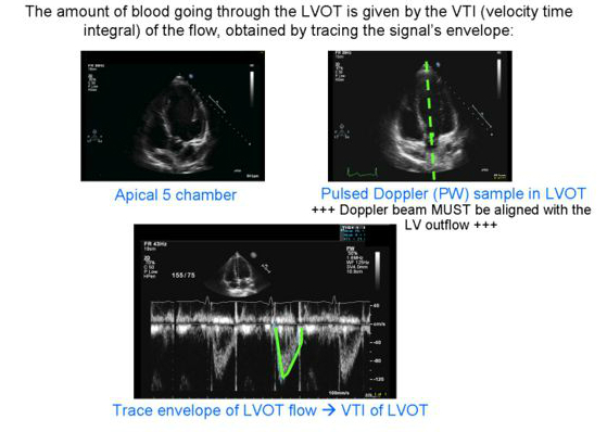
- Calculate CO on echo easily: need LVOT VTI and LVOT diameter (which is normally equal to the BSA! Except in the morbidly obese…)
- SV= Area of LVOT (πR2) x VTI
- Thus plug that into: CO= HR x SV
(Provided by https://web.stanford.edu/)
- When measuring VTI, know your machine. Some are better than others, and a poor machine will underestimate the VTI (normal is 20-25, <20 is considered low)
- Your VTI measurement will confirm your estimation of the EF
- i.e. if your estimation of the EF was low and the VTI is normal, the EF is likely normal for that patient
[/x_text]
[x_text]Suggested Reading
- Maeder MT, Karapanagiotidis S, Dewar EM, Gamboni SE, Htun N, Kaye DM. Accuracy of Doppler echocardiography to estimate key hemodynamic variables in subjects with normal left ventricular ejection fraction.J Card Fail. 2011 May;17(5):405-12. [PubMed Link]
- Borges AC, Kivelitz D, Walde T, Reibis RK, Grohmann A, Panda A, Wernecke KD, Rutsch W, Hamm B, Baumann G. Apical tissue tracking echocardiography for characterization of regional left ventricular function: comparison with magnetic resonance imaging in patients after myocardial infarction.J Am Soc Echocardiogr. 2003 Mar;16(3):254-62.[PubMed Link]
- Price S, Nicol E, Gibson DG, Evans TW. Echocardiography in the critically ill: current and potential roles. Intensive Care Med. 2006 Jan;32(1):48-59. [PubMed Link]
[/x_text]


