Dr. Haase is a man of many talents. Trained in emergency medicine, surgical critical care, critical care medicine, and understands the value of Point of Care Ultrasound (POCUS). This week he’s addressing the often under appreciated and under recognized importance of RV dysfunction. Done a TAPSE of the RV lately? If not, take notes and make sure not to forget the RV.
Review and summary by Dr. Rabin Shrestha
- Anatomical considerations of the RV
- RV contracts mainly by the longitudinal movement (unlike the left ventricle)
- Other movements – RV free wall movement and LV torsion/twisting
Assessment
- RV diastolic function
- View: Assess the RV size and shape with the apical 4 chamber view. Should be a thin walled structure, and crescent in shape
- RVEDA:LVEDA (RV and LV end diastolic areas) – The RV should be < 1/2 the volume of the LV – usually about 1/3 the size
- Tissue Doppler assessment
- Location: Tricuspid annulus
- Checking your E’/a’can help differentiate between acute/chronic RV dysfunction.
- RV systolic function
- First assess with visual inspection and estimation: Septal dyskinesia, septal flattening, or late systolic septal movement are all signs of RV systolic dysfunction.
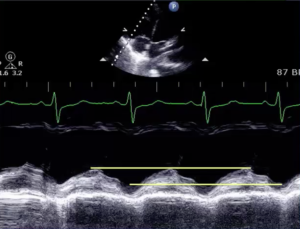
- Tricuspid annular plane systolic excursion (TAPSE) is most useful
- View: Apical 4 chamber view
- Measurement: Obtained by placing the M-Mode cursor through the tricuspid annular plane, then measuring the peak to trough distance.
- Other possible measurements (less useful)
- Tissue Doppler imaging – goal to measure the velocity of the tricuspid annulus (S’)
- Right ventricular fractional area change (RVFAC) is analogous to the ejection fraction of the LV
- Acute vs Chronic
- No definitive criteria to distinguish acute from chronic right heart pathology
- Right ventricular free wall thickness is found in chronic RV strain but RV wall thickening can occur at 48 hours.
- McConnell sign – apical sparing in RV dysfunction – is a sign of acute PE.
Suggested Readings
- Rudski LG, Lai WW, Afilalo J, et al. Guidelines for the echocardiographic assessment of the right heart in adults: a report from the American Society of Echocardiography endorsed by the European Association of Echocardiography, a registered branch of the European Society of Cardiology, and the Canadian Society of Echocardiography. J Am Soc Echocardiogr. 2010;23(7):685-713. [Open Access pdf]
- American Society of Echocardiography – http://asecho.org/ Lots of Open Access Educational Material.


